Clinicians evaluating platelet-rich plasma (PRP) systems often start with what looks like a simple question: should I use a single-spin or a double-spin protocol? The honest answer is that neither is inherently better. Every PRP system is different. The variables that actually shape what gets injected into your patient (and how reproducibly you can produce it) are the volume of blood drawn, the patient’s baseline platelet count, the recovery rate of the specific kit, the leukocyte content, the red blood cell contamination, and the activation method. The spin method is a workflow choice that interacts with all of these. This guide walks through what PRP preparation actually involves, why “single-spin versus double-spin” is not the right framing, and what variables you should actually be tracking in your own practice.
TLDR: PRP preparation is not a single technique. It is a family of techniques that differ across multiple variables: centrifugation method, blood volume drawn, anticoagulant choice, leukocyte content, red blood cell contamination, activation method, and the recovery rate of the specific kit. Neither single-spin nor double-spin is inherently better. Every PRP system is different. Single-spin systems are usually simpler to operate and often use a separator gel that automates component separation. Double-spin systems involve more handling steps and offer different flexibility. Total platelet dose, the variable most strongly linked to outcomes, is essentially the product of the patient’s baseline platelet count, the volume of blood drawn, and the kit’s recovery rate. Recovery rate is the system-level metric that most directly determines what your patient receives. Outcomes vary in every patient.
Important Disclaimer
Regenerative Medicine Academy (RMA) is an education company providing training for licensed clinicians. This article is educational content and does not constitute medical advice, legal counsel, or any guarantee of clinical outcomes. All PRP injection applications discussed are off-label uses of FDA-cleared devices. This article does not provide step-by-step preparation recipes; specific device parameters should be learned from manufacturer instructions for use (IFUs), published studies, and hands-on supervised training. Clinicians are responsible for understanding FDA regulatory status, scope of practice, informed consent, and malpractice implications before implementing any technique. Individual clinical judgment and patient-specific factors must guide all clinical decisions.
If you have attended even one PRP training course, you have probably heard two experienced clinicians express directly opposite preferences about preparation. One swears by single-spin because it is fast, simple, and minimizes handling. The other swears by double-spin because they believe it yields higher platelet counts. Both can be right within their own systems, and both can be wrong if they assume their preferred method is universally superior. The question “single or double” is only a fragment of what actually matters: what is the final platelet dose, leukocyte content, and red blood cell contamination of the product you are injecting, and how reproducibly can you produce it across patients?
This article is for clinicians who want to move past preparation preferences and understand what the variables actually are, how they interact, and where the evidence is strong enough to guide decisions. We will not provide specific centrifuge parameters, volumes, or timings, for two reasons. First, those details are device-specific and properly learned from manufacturer instructions for use and supervised training. Second, the literature does not support naming any single protocol as universally optimal. What we will do is give you the conceptual framework to evaluate any preparation system on its own terms and to read the PRP literature with appropriate skepticism.
What PRP preparation actually involves
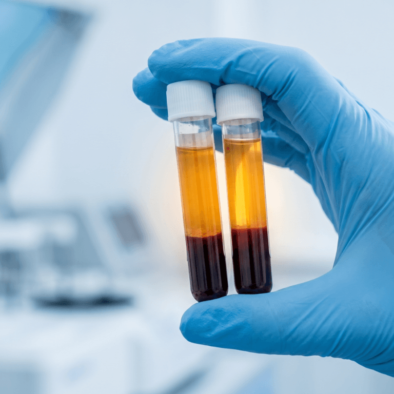
Platelet-rich plasma is an autologous blood product, meaning it comes from the patient’s own venous blood. The core process is simple to describe: draw blood, add an anticoagulant, centrifuge to separate components by density, collect the platelet-enriched fraction, and inject it. The simplicity of that description is misleading, because each step involves choices that independently affect what ends up in the syringe.
The preparation process involves several distinct variables. Blood collection volume typically ranges from 10 to 60 mL drawn before processing, and larger volumes generally yield more total platelets and growth factors. Anticoagulant type determines how well platelets are preserved during processing. The most studied options are acid citrate dextrose solution A (ACD-A), sodium citrate, ethylenediaminetetraacetic acid (EDTA), and heparin. Centrifugation method (single-spin or double-spin) and centrifugation parameters (speed, force measured in relative centrifugal force or RCF, duration, and temperature) determine how components separate. Leukocyte content is the choice between leukocyte-rich PRP (LR-PRP, which retains white blood cells) and leukocyte-poor PRP (LP-PRP, which minimizes them). Red blood cell (RBC) contamination reflects how cleanly platelets are separated from red cells. Activation method is whether platelets are activated before injection using exogenous agents or left to activate inside the body. Final volume and platelet dose determine how much of the product and how many total platelets reach the target tissue.
A separate, often overlooked variable belongs on this list: the kit’s recovery rate. Recovery rate is the percentage of platelets in the original whole blood sample that the system actually captures into the final product. It varies meaningfully between commercial kits and is one of the most important system-level metrics, because it sits at the intersection of every other preparation choice. We will return to this later.
No single combination of these variables is universally optimal. Each interacts with the others, and the right combination depends on the clinical indication, the anatomic target, the device, and patient biology. A useful way to think about preparation is that your centrifuge does not produce PRP. It produces a specific PRP with a specific composition, and different centrifuges and different kits produce different PRPs.
What this means in practice: When clinicians say “I use PRP for knee osteoarthritis,” the critical missing information is which PRP, prepared with what anticoagulant, at what parameters, with what leukocyte content, at what blood volume, with what kit recovery rate, and at what final platelet dose. Two clinicians answering the same question can be describing fundamentally different products. That ambiguity matters both for your own outcomes and for how you interpret the literature.
Single-spin versus double-spin: the mechanical differences
Centrifugation is the heart of PRP preparation because it is the step that separates blood components by density. When whole blood is spun, the heaviest components (red blood cells) settle at the bottom, platelets and white blood cells form a thin layer called the buffy coat in the middle, and the plasma stays on top. The choice between single-spin and double-spin protocols is a choice about how to capture and concentrate the platelet-containing layer.
Before describing each method in detail, the most important point: every PRP system is different. The idea that double-spin is automatically better than single-spin, or vice versa, does not hold up. Yields, recovery rates, leukocyte content, and red blood cell contamination all vary kit by kit. A well-designed single-spin kit can produce a comparable or better product than a poorly executed double-spin protocol that loses platelets in the transfer step. The spin method tells you something about the workflow and the handling steps. It does not tell you what the patient will actually receive.
How single-spin systems work
In a single-spin protocol, blood undergoes one centrifuge cycle. The cycle separates the heavy red blood cells from the upper plasma layer, which contains platelets and, depending on parameters, varying amounts of white blood cells. The clinician then aspirates the platelet-rich upper layer.
Many modern single-spin kits include a separator gel inside the collection tube. The gel has a density that falls between the red blood cells (heavy) and the platelet-plasma layer (lighter). During centrifugation, the gel migrates to position itself between the two layers, forming a physical barrier. After the spin, the clinician can aspirate or pour off the platelet-rich plasma cleanly above the gel without disturbing the red blood cells trapped below it. This design eliminates the transfer step that double-spin protocols require, reduces the chance of red blood cell contamination, and makes the overall workflow simpler and more reproducible.
Single-spin systems are usually simpler to operate. There are fewer handling steps, less opportunity for premature platelet activation during transfer, and a shorter total preparation time. For a busy clinic, those operational advantages are real. The tradeoff is that single-spin kits can have less flexibility to fine-tune leukocyte content, and final platelet concentration depends heavily on the specific gel formulation, tube design, and recommended centrifuge parameters of the particular kit. The right way to think about a single-spin system is as an integrated package: the tube, the gel, the recommended spin parameters, and the recovery rate are designed together, and changing any one of them in isolation can change the product.
How double-spin protocols work
In a double-spin protocol, blood undergoes two sequential centrifuge cycles. The first spin separates red blood cells from the plasma layer. The clinician then transfers the plasma layer to a second tube, where a second spin concentrates the platelets into a pellet at the bottom. The platelet-poor plasma above that pellet is either discarded or partially retained, and the pellet is resuspended in a small volume of plasma to create the final product.
Double-spin protocols can offer more flexibility to manipulate leukocyte content and final concentration, and they often allow a smaller final volume. Some clinicians and researchers prefer double-spin for that reason in specific applications. The tradeoffs are also real: additional handling creates opportunities for premature platelet activation, the transfer step creates a risk of red blood cell contamination if pipetting is imprecise, preparation time is longer, and operator variability is higher because there are more steps where technique affects outcome. A poorly executed double-spin protocol can lose substantial platelets in the transfer step, which lowers the system’s effective recovery rate.
What the literature actually shows
Comparative studies of single-spin and double-spin protocols give mixed results, which is exactly what you would expect if the spin method itself is not the dominant variable. Some studies find double-spin yields higher concentrations on average. A direct comparison study found that a manual double-spin method achieved a mean platelet count approximately 4.8 times higher than a commercially available automated single-step device, with statistically significantly higher platelet capture efficiency (47.1% versus 31.9%). An 18-protocol evaluation by Perez and colleagues that tested both techniques reported that double-spin methods concentrated platelets to a greater degree overall, though outcomes varied markedly across different parameter combinations within each category.
Other studies find the opposite. A randomized, double-blind clinical trial in dermatology found that a single-spin protocol produced a 5.81-fold increase in platelet count, exceeding the double-spin method tested in the same study. This kind of finding is not surprising once you accept that the number of spins is not the dominant variable. Specific parameters (force, time, blood volume, anticoagulant, gel design, operator technique) matter more than whether one or two spins are performed.
What this means in practice: Comparing “single-spin” and “double-spin” as categories is not the right question. The right question is how much total platelet dose each specific kit reliably delivers in your hands, in your patient population, with your blood draw volumes. Spin method is a workflow choice. Recovery rate, blood volume, and patient factors are what determine the final dose.
Why platelet dose matters more than spin method alone
The most clinically useful way to think about preparation is through the lens of total platelet dose delivered to the target tissue. Recent evidence increasingly supports the idea that platelet dose, not concentration factor or spin method alone, drives clinical outcomes. This reframing matters because it tells you what to optimize when you are evaluating a preparation system or troubleshooting inconsistent outcomes in your practice.
A 2024 systematic review in Arthroscopy specifically evaluated platelet dose across PRP injection studies for musculoskeletal conditions. Among 29 studies analyzed, the 28 studies that reported statistically significant positive outcomes at 6 months had a mean platelet dose of approximately 5,500 × 10^6 platelets per injection, while the single study that did not report a positive outcome used a lower dose. A 2025 meta-analysis published in the American Journal of Sports Medicine by Bensa and colleagues found that higher platelet concentrations correlated with greater knee osteoarthritis benefit, supporting a threshold-effect framework rather than a one-size-fits-all concentration target. For context on scale, 1 billion platelets is 1,000 × 10^6 platelets.
The dose framework is particularly well supported for lateral epicondylitis. A 2025 systematic review and meta-analysis of randomized controlled trials found that studies using high-dose PRP (defined as greater than a 3-fold concentration increase over whole blood) produced significantly better visual analog scale outcomes than studies using low-dose PRP, with a mean difference of -1.31 favoring the high-dose group.
Recovery rate: the system-level metric that matters most
Recovery rate is the percentage of platelets in the original whole blood sample that end up in the final PRP product. It is a direct measure of how efficiently a specific kit captures platelets, and it varies meaningfully between commercial systems. A kit with a 70 percent recovery rate captures most of the available platelets; a kit with a 30 percent recovery rate loses most of them. This single variable can matter more than the concentration factor or the spin method.
The math is straightforward. Total platelets in the final product is essentially the product of three numbers: the patient’s baseline platelet count, the volume of blood drawn, and the kit’s recovery rate. Consider a patient with a normal baseline platelet count of 250,000 platelets per microliter. With a 30 mL blood draw and a kit that has an 80 percent recovery rate, the final product contains roughly 6 billion platelets. The same patient with the same blood draw using a kit with a 30 percent recovery rate ends up with roughly 2.25 billion platelets. The difference between those two outcomes is not the spin method. It is the recovery rate.
Drawing more blood is a related lever. If you draw 30 mL instead of 15 mL, you are starting with twice as many platelets. The same recovery rate then captures twice as many platelets in absolute terms. This is why blood draw volume varies across protocols, and why some advanced applications use larger draws to support higher cumulative doses. More blood drawn generally means more platelets and more growth factors collected, assuming the kit’s recovery rate stays consistent across volumes.
The relationship between concentration factor and total dose is also worth understanding clearly. A single-spin protocol that produces a 4-fold concentration and is injected at a larger volume can deliver an equivalent or even greater total platelet dose than a double-spin protocol that produces a 7-fold concentration but is injected at a smaller volume. Concentration factor by itself does not tell you total dose. You also need to know the volume.
What this means in practice: When evaluating a PRP kit, the question worth asking the manufacturer is what the recovery rate is in published or internal data, not just what the concentration factor is. When evaluating your own outcomes, the metric to track is total platelet dose per injection, calculated from your hemoanalyzer-verified platelet count and your injection volume. Recovery rate is the variable that connects what is in the patient’s blood to what is in the syringe.
The leukocyte question: LP-PRP versus LR-PRP
Leukocyte content is the second major preparation variable that shapes clinical decision-making. Most quality PRP training programs teach a working distinction: LP-PRP minimizes white blood cells and may be preferred for intra-articular applications, while LR-PRP retains white blood cells and may be better suited for tendon applications. This distinction is clinically reasonable as a conservative starting point, but the underlying evidence is more nuanced than the simple framing suggests.
The laboratory data is striking. In vitro studies of synoviocytes (the cells lining the joint) show that LR-PRP causes approximately 4.9 percent synoviocyte death, compared with 0.72 percent with LP-PRP. That is nearly a 7-fold difference in a laboratory model. RBC concentrate, for comparison, causes 12.5 percent synoviocyte death in the same model. These findings form the historical rationale for preferring LP-PRP in intra-articular applications, where chondrocyte (cartilage cell) and synovial preservation are clinical priorities.
The clinical data tells a more complicated story. A 2026 systematic review and meta-analysis specifically comparing LR-PRP and LP-PRP for knee osteoarthritis concluded that current evidence is insufficient to determine whether leukocyte presence is beneficial or detrimental in that clinical context. The review found no statistically significant differences in pain reduction or functional improvement between the two groups across included randomized controlled trials. Some newer data even suggests LR-PRP may express higher levels of IL-1Ra (interleukin-1 receptor antagonist, an anti-inflammatory protein) and IL-4 in knee osteoarthritis patients, hinting at potential anti-inflammatory effects in chronic low-grade inflammation contexts that the in vitro toxicity data would not predict.
The 2024 ESSKA-ORBIT consensus on PRP for knee osteoarthritis takes this head-on. The consensus group considers both LP-PRP and LR-PRP as valid options for the management of knee osteoarthritis when PRP is being considered, with the reasoning that effectiveness is likely multifactorial and the dependence on leukocyte presence alone may be overestimated.
What this means in practice: LP-PRP has been associated with reduced inflammatory cytokine production in laboratory models, but current clinical evidence has not demonstrated significant differences in patient outcomes between LP-PRP and LR-PRP preparations for osteoarthritis. The traditional framework of LR-for-tendons and LP-for-joints remains a reasonable conservative default, but clinicians should understand that it is based on in vitro data and theoretical concern rather than head-to-head clinical trials showing a definitive outcome difference.
Other preparation variables that shape the final product
Beyond spin method, recovery rate, and leukocyte content, three other variables materially affect what ends up in the syringe: anticoagulant choice, activation method, and red blood cell contamination. Each independently shapes product biology.
Anticoagulant choice
The anticoagulant added to the collection tube prevents the blood from clotting during processing. Acid citrate dextrose solution A (ACD-A) is the most widely preferred anticoagulant for PRP intended for musculoskeletal use. It works by chelating ionized calcium, a process that binds calcium and prevents the coagulation cascade from starting. ACD-A is considered superior to simple sodium citrate and heparin for preserving platelet structural integrity and downstream growth factor release. PRP prepared with ACD-A has been shown to release significantly more transforming growth factor beta-1 (TGF-beta1) than preparations using heparin or simple citrate.
ACD-A has one important caveat: its acidifying effect lowers blood pH to approximately 6.5 during processing. Some protocols recommend buffering the PRP back toward physiologic pH before injection, though this is not universally practiced. Heparin is generally not recommended for PRP intended for regenerative injection because it can directly interfere with platelet function and may inhibit growth factor release through interactions with platelet surface proteins. EDTA is similarly avoided because it can trigger platelet activation during collection, releasing growth factors prematurely.
Activation method
PRP can be injected in its resting (unactivated) state, relying on the patient’s own tissue collagen and other signals to trigger platelet degranulation inside the body, or it can be activated before injection using exogenous agents. Four activation methods appear in the literature most often: calcium chloride, autologous thrombin, a combination of both, and collagen type I.
A 2016 review noted that the choice of strategy to activate PRP is mainly based on practical reasons rather than supported by studies on the effects of the different procedures on the final platelet releasate. That observation remains largely true today. The differences between activation methods are primarily in growth factor release kinetics rather than total growth factor output. Calcium chloride produces a more progressive, sustained release over time. Thrombin and the combination produce more immediate release. Collagen type I produces the lowest overall growth factor release of the four. In situ activation (no pre-activation, allowing tissue signals inside the body to trigger release) is commonly preferred for musculoskeletal injections because it avoids premature growth factor release during preparation and delivery.
Red blood cell contamination
Red blood cell contamination deserves its own attention, particularly for intra-articular use. A 2023 editorial in Biomedicines titled “Red Blood Cells in Platelet-Rich Plasma: Avoid If at All Possible” summarized evidence that RBC breakdown releases hemoglobin, iron, and hemin, which can activate inflammatory pathways, generate oxidative stress, and contribute to chondrocyte and synovial cell damage. Both in vitro and animal model data link RBC exposure to cell death and long-term degenerative changes. Single-spin kits with separator gel and well-executed double-spin protocols can both achieve low final hematocrits. The kit design, the gel formulation, and the operator’s technique all affect how cleanly platelets separate from red cells.
What this means in practice: Anticoagulant choice and RBC minimization are two preparation levers that do not require specialized equipment to optimize, yet are frequently overlooked. If you are using a commercial PRP kit, know which anticoagulant ships with it and what the typical final hematocrit is. These are preparation details that matter for your product’s biology.
Classification systems: why “PRP” is not one product
Five major classification systems exist to describe and categorize PRP preparations. None has been validated as a predictor of clinical outcomes, but each captures something meaningful about the heterogeneity of products under the PRP label. Understanding them matters both for interpreting the published literature and for communicating accurately about your own protocols.
The classification systems use different criteria. Ehrenfest stands for the Ehrenfest 2009 classification. PAW stands for platelet, activation, white blood cell (the DeLong 2012 system). PLRA stands for platelet, leukocyte, red blood cell, activation (the Mautner 2015 system). DEPA stands for dose, efficiency, purity, activation (the Magalon 2016 system). MARSPILL stands for method, activation, red blood cell, spin, platelet, image guidance, light activation, leukocyte (the 2017 Lana system). In the following table, P-PRP means pure platelet-rich plasma, L-PRP means leukocyte platelet-rich plasma, P-PRF means pure platelet-rich fibrin, and L-PRF means leukocyte and platelet-rich fibrin.
| System | Year | Key criteria | Output format |
| Ehrenfest | 2009 | Leukocyte content, fibrin architecture | Four categories (P-PRP, L-PRP, P-PRF, L-PRF) |
| PAW (DeLong) | 2012 | Platelet count, activation, white cell presence | Three-variable code |
| PLRA (Mautner) | 2015 | Platelet concentration, leukocytes, RBCs, activation | Four-variable code |
| DEPA (Magalon) | 2016 | Dose, efficiency, purity, activation | Letter grade (A through D) |
| MARSPILL | 2017 | Eight variables including spin method | Eight-variable code |
The DEPA system is particularly interesting because it explicitly includes both dose and efficiency (a close cousin of recovery rate) as graded variables. The MARSPILL system explicitly includes spin method as a dimension. The deeper message across all five systems is the same: when you read a PRP study, the word “PRP” by itself is nearly empty as a description of the intervention. Two studies using diametrically different preparations can both report “PRP” as their treatment arm. The classification systems exist precisely because the field has recognized this heterogeneity problem and has been trying, for over 15 years, to develop a common language.
What this means in practice: When evaluating a new PRP study or a device claim, ask which classification the authors or manufacturers use, what their final product composition actually is, and whether the preparation matches what you are doing in your own practice. A positive result in a study using a DEPA grade A preparation tells you little about the performance of a preparation that would score a grade C or D under the same system.
The standardization crisis and what it means for your practice
The most important context for any discussion of PRP preparation is the scale of the standardization problem in the field. A landmark analysis published in the Journal of Bone and Joint Surgery in 2017 found that 84 percent of PRP clinical studies did not provide quantitative metrics on the composition of the final PRP product, and the majority did not include sufficient information to reproduce the protocol. This is not a minor reporting gap. It means that most of the PRP evidence base cannot be meaningfully interpreted with respect to what was actually injected.
The Minimum Information for Studies Evaluating Biologics in Orthopaedics (MIBO) guidelines, also published in 2017, established minimum reporting requirements for PRP clinical studies. The checklist asks authors to report the details that would let other clinicians and researchers understand and reproduce the intervention: blood volume collected, anticoagulant used, centrifuge parameters, final platelet count, leukocyte content, RBC content, activation method, volume injected, and more. Despite being published nearly a decade ago, adherence remains poor. A systematic review published in January 2026 confirmed persistently poor MIBO adherence across the broader field.
This is why a high-profile negative trial like the 2021 RESTORE trial in JAMA is difficult to generalize from. RESTORE found that PRP was no better than saline at 12 months for knee osteoarthritis, but interpreting that finding for your own practice requires knowing how the RESTORE preparation compared to whatever kit you use. Without standardized reporting, this comparison is largely guesswork.
The AAOS Orthobiologics Registry, announced on December 5, 2025, directly addresses this gap. The registry is designed to measure the long-term safety, efficacy, and real-world outcomes of orthobiologic therapies in patients with knee osteoarthritis, beginning with a pilot of 10 participating sites and plans for methodical expansion over three to five years. What makes the registry structurally different from prior PRP data collection efforts is its incorporation of a standardized hemoanalyzer configuration, which together with biorepository linkage will allow the actual composition of injected PRP to be tracked alongside patient-reported outcomes.
Even commercial PRP kits do not always deliver what their manufacturers report. A retrospective analysis of 118 PRP preparations using a commonly used commercial kit during routine clinical care found that while most preparations met or exceeded manufacturer platelet enrichment expectations, modification of yield volume significantly affected platelet enrichment ratios, and patient age and gender both moderately affected platelet fold enrichment from baseline. This means that clinicians who rely solely on manufacturer-reported yield specifications, without point-of-care verification, may have incomplete knowledge of what they are actually injecting. The kit’s stated recovery rate is an average across patients, not a guarantee for any individual patient.
What this means in practice: The standardization crisis is not an academic concern. It directly affects how you should interpret published outcomes, how you should evaluate commercial kits, and how you should think about the consistency of your own practice. Point-of-care platelet counting, whether through a portable hemoanalyzer or a laboratory send-out, is the only way to know what dose you are actually delivering to each patient. Until MIBO-compliant reporting becomes the norm in published literature, reasonable skepticism about any claimed PRP outcome remains warranted.
A fictional day-in-the-lab perspective
The following vignette is a fictional composite created for educational purposes. It does not represent a real clinician.
Dr. Chen is a sports medicine physician who has been offering PRP in her practice for about 18 months. She has noticed something that bothers her: her outcomes seem inconsistent. Some patients respond beautifully, others do not respond at all, and she cannot clearly predict which will be which. She decides to attend a hands-on advanced PRP course to understand what might be driving the variability.
During the preparation module, the instructor asks each attendee to describe their current setup. Dr. Chen describes her kit, which is a commercial single-spin system with a separator gel that she chose primarily because a device representative demonstrated it and the protocol seemed simple. She has not been tracking final platelet counts because she assumed the manufacturer’s stated yield was reliable.
The instructor does not tell her she has been doing it wrong. The instructor’s point is not that she should switch to double-spin. It is that her kit, like any system, has a specific recovery rate, a specific blood draw volume, and a specific set of recommended parameters that together determine what she ends up injecting. She has been operating without verifying her actual platelet yield, which means she has been operating without knowing her actual dose. Two patients of identical size with similar diagnoses could have been receiving meaningfully different doses, not because of her technique but because of patient-level variability in baseline platelet count, blood volume drawn, and the kit’s variable capture efficiency from patient to patient.
Dr. Chen spends the afternoon lab session learning more about her own kit and observing how a manual double-spin technique compares. Once she sees the visible layering after each spin and watches how the separator gel in her own kit positions itself, she has a much clearer mental model of what her system is doing each time she runs it. The instructor demonstrates point-of-care hemoanalyzer verification and explains that this is the single change that would most improve her ability to track her own outcomes. If she knows her final platelet count for each patient, she can start correlating her outcomes with her actual doses rather than with the manufacturer’s theoretical yield.
The real lesson for Dr. Chen is not that single-spin or double-spin is better. It is that she has been treating preparation as a fixed step in the protocol when it is actually a variable that changes with every patient. She leaves the course intending to add hemoanalyzer verification before her next injection, to start recording final platelet counts in her chart, and to revisit her approach to musculoskeletal regenerative medicine training once she has three months of verified data.
Patient selection and what this means for your practice
None of the preparation variables we have discussed matter if the patient is not an appropriate candidate for PRP in the first place. Patient selection remains the single largest determinant of outcome, and no preparation approach compensates for inappropriate selection. That said, a few practical connections between preparation and clinical decision-making are worth keeping in mind.
For intra-articular applications such as knee osteoarthritis, the considerations that tend to matter most are minimizing red blood cell contamination, using LP-PRP as a conservative default (recognizing that clinical evidence does not show superiority over LR-PRP for osteoarthritis outcomes), and ensuring adequate total platelet dose. For tendon applications such as lateral epicondylitis or plantar fasciitis, some clinicians prefer LR-PRP based on the theoretical rationale that a modest inflammatory response may support tendon remodeling, though again the comparative clinical evidence is limited. For all applications, platelet dose matters, and the recent literature suggests a threshold effect rather than a linear dose-response.
Patient factors also influence what you can achieve with any given preparation system. Baseline platelet count varies substantially between patients. A patient with a lower baseline who undergoes the same protocol will end up with a lower final dose than a patient with a higher baseline. Hematocrit affects how cleanly platelets separate during centrifugation. Recent NSAID (nonsteroidal anti-inflammatory drug) use, corticosteroid exposure, and certain comorbidities all affect platelet function beyond what a centrifuge can influence. These patient-level factors are reasons why following a standardized preparation protocol does not always produce a standardized product.
The practical path to consistency involves three elements: learning one or two preparation systems well through hands-on supervised training rather than dabbling across many, verifying your platelet yield for each patient rather than relying on manufacturer-reported specifications, and tracking your outcomes systematically so that you can correlate real-world doses with real-world results over time. RMA’s online regenerative medicine education covers the conceptual foundation, and hands-on courses build the procedural and verification habits that translate concept into reproducible practice.
Frequently asked questions
Is single-spin or double-spin better?
Neither is universally better. Every PRP system is different, and the spin method tells you about workflow rather than about clinical performance. Single-spin systems are usually simpler to operate, often use a separator gel that automates component separation, and have fewer handling steps. Double-spin systems offer more flexibility to manipulate leukocyte content and concentration but require more handling. Recovery rate, the percentage of available platelets that end up in the final product, varies meaningfully between specific kits and matters more than the spin method itself. A well-designed single-spin kit can deliver more total platelets to the patient than a poorly executed double-spin protocol that loses platelets in the transfer step.
What is recovery rate, and why does it matter?
Recovery rate is the percentage of platelets in the original whole blood sample that end up in the final PRP product. It varies meaningfully between commercial kits. Total platelets in the final product is essentially the product of three numbers: the patient’s baseline platelet count, the volume of blood drawn, and the kit’s recovery rate. A kit with a high recovery rate captures most of the available platelets; a kit with a low recovery rate loses most of them. Recovery rate is the system-level metric that most directly determines what your patient receives.
Does drawing more blood produce more platelets?
Yes, generally. If you draw 30 mL instead of 15 mL, you are starting with twice as many platelets in absolute terms. Assuming the kit’s recovery rate stays consistent across volumes, the same recovery rate captures twice as many platelets. This is why blood draw volume varies across protocols and why some applications use larger draws to support higher cumulative doses. Drawing more blood is a practical lever for total dose, separate from the spin method.
What platelet concentration should I target?
No validated universal concentration has been established. The commonly cited “5 times baseline” figure is a rough average from the older literature, not a validated clinical threshold. More recent work suggests a dose threshold near 3.5 billion platelets per injection for clinical benefit, with cumulative course doses of 10 to 12 billion platelets associated with optimal outcomes in knee osteoarthritis and tendinopathy studies. The practical target depends on your specific kit, patient population, and indication.
Do I need to measure my platelet yield?
If you are not measuring your final platelet count, you do not actually know what dose you are delivering. Manufacturer-reported yields are averages and do not account for patient-level variability in baseline platelet count, blood volume drawn, hematocrit, or system capture efficiency. Point-of-care hemoanalyzer verification is the most reliable way to know what is in the syringe before injection, and it is the single change most likely to improve your ability to track and interpret your own outcomes.
Which anticoagulant should I use?
Acid citrate dextrose solution A (ACD-A) is the most widely preferred anticoagulant for PRP intended for musculoskeletal use because it preserves platelet integrity and supports downstream growth factor release. Heparin is generally not recommended because it can interfere with platelet function. EDTA is generally avoided because it can trigger premature platelet activation during collection. Your specific commercial kit may ship with a specific anticoagulant. Know what it is and why.
Should I activate PRP before injection or let it activate in situ?
In situ activation (no pre-activation) is commonly preferred for musculoskeletal injections because it avoids premature growth factor release during preparation and delivery. The comparative clinical evidence for activation versus no activation is limited, and no consensus exists on optimal activation method by indication.
Is LP-PRP really better than LR-PRP for joints?
The laboratory data show significantly less synoviocyte death with LP-PRP (0.72 percent) than with LR-PRP (4.9 percent), which forms the theoretical rationale for preferring LP-PRP in intra-articular applications. The clinical data, however, do not show significant differences in patient outcomes between LP-PRP and LR-PRP for knee osteoarthritis in a 2026 systematic review and meta-analysis. LP-PRP remains a reasonable conservative default for joints, but the idea that LR-PRP is actively harmful in the joint is not supported by current clinical evidence.
Where can I learn device-specific preparation protocols?
Device-specific protocols should come from manufacturer instructions for use, published studies using the same system, and hands-on supervised training. This article deliberately does not provide specific centrifuge parameters, volumes, or timings because those details are properly device-specific and are best learned in a supervised clinical setting. Online regenerative medicine education and in-person courses are designed to cover the specific protocols that match the systems you will use in your practice.
Key takeaways
- PRP preparation involves multiple interdependent variables (blood volume drawn, anticoagulant, centrifugation method and parameters, leukocyte content, red blood cell contamination, activation method, kit recovery rate, and final platelet dose). The single-spin versus double-spin distinction is only one of them and is not the most clinically important.
- Neither single-spin nor double-spin is inherently better. Every PRP system is different. Single-spin systems are usually simpler to operate and often use a separator gel that automates component separation. Double-spin systems offer different flexibility but require more handling.
- Total platelet dose delivered to the patient is essentially the product of three numbers: the patient’s baseline platelet count, the volume of blood drawn, and the kit’s recovery rate. Drawing more blood generally yields more platelets and growth factors. Recovery rate is the system-level metric that most directly affects what your patient receives.
- LP-PRP shows substantially less synoviocyte toxicity than LR-PRP in laboratory models, but 2026 clinical evidence does not show outcome differences between the two for knee osteoarthritis. The ESSKA-ORBIT 2024 consensus considers both valid options.
- Five major classification systems (Ehrenfest, PAW, PLRA, DEPA, MARSPILL) exist to describe PRP preparations. None has been validated as an outcome predictor, but they underscore that “PRP” by itself is an incomplete description of any intervention.
- The standardization crisis is real and clinically relevant. Most published PRP studies do not report enough preparation detail to be reproduced, and point-of-care hemoanalyzer verification is currently the most reliable way to know what you are actually injecting in your own practice.
- Device-specific preparation protocols should come from manufacturer instructions for use, published studies using the same system, and hands-on supervised training. Articles like this one can provide a conceptual framework but should not be used as recipe substitutes.
About Regenerative Medicine Academy
Regenerative Medicine Academy (RMA) is a medical education company providing hands-on, small-group regenerative medicine training courses for licensed healthcare professionals. RMA does not provide clinical treatments or medical services. All course content is educational and designed to support clinicians in making informed, evidence-based decisions within their scope of practice. For information on upcoming training dates, visit regenmedacademy.com.
Off-label use note
Procedures discussed in this article involve off-label use of FDA-cleared devices. PRP preparation devices are cleared through the FDA’s 510(k) pathway for producing platelet-rich preparations to mix with bone graft materials in orthopedic surgical procedures. All intra-articular, peritendinous, and other musculoskeletal injection applications are off-label. Clinicians may use FDA-cleared devices off-label based on clinical judgment but must obtain informed consent, discuss evidence, risks, and alternatives, and document thoroughly.
Platform and jurisdiction note
Scope of practice, supervision requirements, and corporate practice of medicine rules vary by state. Clinicians should consult their state medical board, malpractice carrier, and legal counsel regarding compliance in their jurisdiction before offering regenerative procedures.
For clinicians ready to take the next step
Clinicians who want to deepen their understanding of preparation variables, recovery rate verification, and reproducible PRP technique can review RMA’s full course catalog, which covers musculoskeletal, cosmetic, sexual health, and adipose tissue applications. Hands-on training is the practical way to build the system-specific habits that turn the conceptual framework in this article into reproducible clinical practice.