Point-of-care ultrasound has reshaped how clinicians deliver regenerative injections. The evidence consistently shows that imaging guidance places needles more accurately than landmark-based techniques, but the skill takes months to develop and has no single mandatory U.S. credential. This guide walks clinicians through what the data show, where outcomes evidence is strong, where it remains mixed, and what structured training looks like.
TLDR: Ultrasound-guided injections (USGIs) consistently achieve better needle placement accuracy than landmark-guided injections, with the strongest evidence in the shoulder, knee, small joints, and carpal tunnel. For regenerative procedures such as platelet-rich plasma (PRP), which is FDA-cleared only for bone graft handling and used off-label for musculoskeletal and aesthetic indications, accurate intra-target delivery is a prerequisite for any biologic effect. Musculoskeletal ultrasound has a steep learning curve, operator skill drives outcomes, and structured training protects both patient safety and your license. This article covers the evidence, the competencies, and the credentialing pathways clinicians should understand before offering these procedures.
Important Disclaimer
Regenerative Medicine Academy (RMA) is an education company providing training for licensed clinicians. This article is educational content and does not constitute medical advice, legal counsel, or a guarantee of clinical outcomes. Techniques discussed may be used off-label and are subject to state and federal regulations. Clinicians are responsible for understanding FDA status, scope of practice, informed consent, and malpractice implications before implementing any technique in practice. Individual clinical judgment and patient-specific factors must guide all clinical decisions.
A familiar scene in the MSK clinic
If you have watched a colleague place a needle into a shoulder without ultrasound, then repeat the same injection under imaging, the difference is hard to unsee. The first approach relies on palpation and anatomic memory. The second lets you see the needle tip, confirm it sits inside the joint or tendon sheath, and watch the injectate spread in real time.
That second view has become standard in many musculoskeletal and regenerative practices. Point-of-care ultrasound has grown from a diagnostic curiosity into a procedural tool that shapes daily workflow. For regenerative injections in particular, where the biologic only works if it reaches the target tissue, image guidance is increasingly treated as expected rather than optional.
The skills do not arrive with the machine, however. Learning to acquire clean images, interpret sonographic anatomy, track a needle in real time, and avoid nearby vessels and nerves takes supervised practice. The rest of this guide covers what the evidence shows, what safe technique looks like, and what kind of training builds real competence.
Why ultrasound guidance has become central to regenerative medicine
Ultrasound guidance is now recognized by major specialty societies as integral to modern musculoskeletal practice. The American Academy of Physical Medicine and Rehabilitation has stated that diagnostic and interventional MSK ultrasound is a fundamental component of physiatric practice and an essential tool for musculoskeletal care. The American Medical Society for Sports Medicine has published a position statement and a recommended fellowship curriculum that treat interventional ultrasound as a core sports medicine skill.
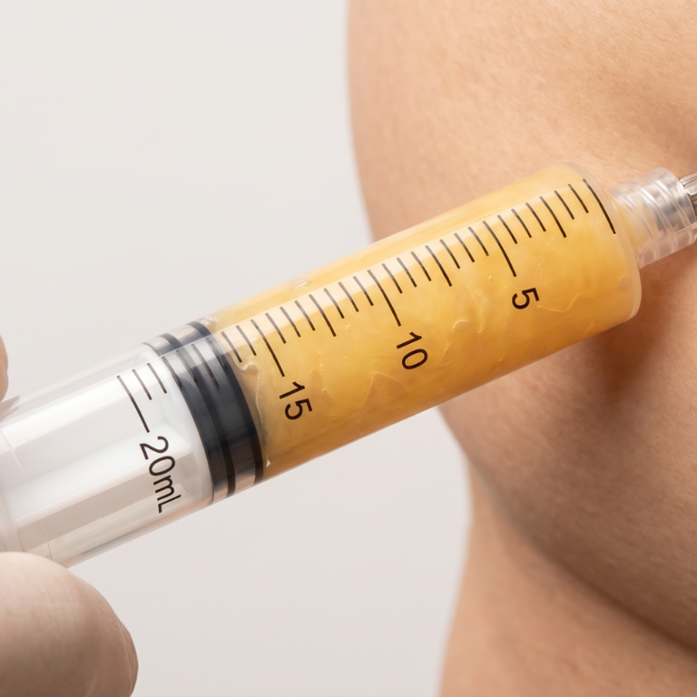
The rationale for regenerative injections is direct. Biologic products such as PRP depend on delivery into the correct compartment. A platelet concentrate that lands in subcutaneous fat rather than an intra-articular space (inside the joint capsule) cannot interact with cartilage or ligament tissue as the clinician intended. Imaging converts the injection from an anatomic estimate into a visible procedure, and it lets you document what was done.
What this means in practice: Ultrasound does not replace clinical reasoning or patient selection. It adds a verification step. You can see what you are injecting, where the fluid travels, and what structures you must avoid.
How ultrasound-guided injections work
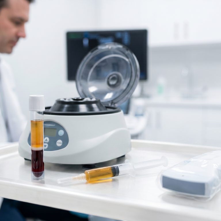
Ultrasound uses high-frequency sound waves to create a real-time image of soft tissue. A probe placed on the skin emits pulses, captures the echoes that return from tissue interfaces, and renders them as grayscale images. Muscles, tendons, ligaments, bursae, nerves, and blood vessels each have characteristic appearances, which lets a trained operator identify the target before the needle enters the skin.
For an injection, the clinician selects a probe suited to the depth of the target. A high-frequency linear probe in the 10 to 15 MHz range works well for superficial structures like small joints or tendons. A lower-frequency curvilinear probe in the 2 to 6 MHz range penetrates deeper tissue such as the hip or lumbar spine. The operator then chooses a needle path. In-plane means the needle shaft runs along the long axis of the probe and stays fully visible throughout the procedure. Out-of-plane means the needle crosses the probe and appears as a bright dot on the screen.
Once the target is identified and the skin is prepped, the clinician advances the needle under continuous imaging. The operator confirms the tip sits where intended, aspirates briefly to rule out intravascular placement, and injects slowly while watching the fluid distribute. Real-time feedback lets the operator reposition if the spread is wrong. That feedback is what separates a guided procedure from a landmark injection, and it is the reason accuracy rises.
The accuracy evidence: ultrasound versus landmark-guided injection
Current research strongly supports ultrasound guidance for needle placement accuracy across most musculoskeletal sites. A 2024 umbrella review published in the Journal of Rehabilitation Medicine synthesized the existing systematic reviews and meta-analyses and concluded that USGIs are more accurate than landmark-guided injections, with particularly consistent findings in the shoulder. Earlier site-specific studies and meta-analyses reinforce the same pattern.
The table below summarizes accuracy data from multiple anatomic sites. Placement accuracy refers to the proportion of injections in which the needle tip was confirmed in the intended target. Clinical outcomes are reported where relevant trials measured pain or function.
| Anatomic site | Ultrasound-guided accuracy | Landmark-guided accuracy | Notes |
| Intra-articular knee | Approximately 95 to 100 percent | Approximately 77 to 83 percent | Level I systematic review; significant difference favoring ultrasound |
| Glenohumeral joint (cadaveric) | About 92 percent | About 72 percent | Cadaveric study; accuracy advantage held regardless of surgeon experience |
| Subacromial space | More accurate with ultrasound | Less accurate with landmark | Meta-analyses also show modest pain relief advantage at 6 weeks |
| Carpal tunnel | Lower nerve irritation rate (around 2 percent) | Higher nerve irritation rate (around 14 percent) | Randomized trial in median nerve injections |
| Small joints (hand, wrist) | Higher accuracy with ultrasound | Lower accuracy with landmark | Consistently reported across reviews |
The effect sizes vary by site and by the specific comparison, and not every randomized trial shows a clinical outcome advantage that matches the accuracy advantage. Still, the accuracy signal is consistent: imaging guidance places needles where they are supposed to go at higher rates than palpation alone, and the difference widens as the target gets smaller or deeper.
What this means in practice: Higher placement accuracy does not translate directly into guaranteed patient improvement. It means the injection reached the intended tissue, which is a necessary condition for any biologic or pharmacologic effect, not a sufficient one.
Where the outcomes evidence is strong, and where it remains mixed
Accuracy and clinical outcomes are related, but they are not the same thing. For regenerative injections in particular, the outcomes literature is more nuanced than the accuracy literature, and honest interpretation matters.
For knee osteoarthritis, a 2025 Bayesian network meta-analysis in Frontiers in Medicine analyzed 14 randomized controlled trials covering 934 patients and compared ultrasound-guided PRP, hyaluronic acid, corticosteroids, ozone, and placebo. PRP ranked highest on several outcome scales in this analysis, including WOMAC pain, stiffness, and function measures. Those rankings reflect the probability that PRP occupied the best rank across simulations, not a confirmed statistical superiority. Most pairwise WOMAC comparisons between active agents did not reach statistical significance, and confidence intervals were wide. The authors also noted that heterogeneity in PRP preparation across studies was substantial. Platelet concentration, leukocyte content, and activation method all vary widely, and those differences likely influence which patients respond. Clinicians who want a deeper look at PRP evidence and technique can also review related coverage on RMA’s regenerative medicine blog.
For subacromial impingement, a 2022 meta-analysis by Deng and colleagues pooled 12 randomized controlled trials covering 891 patients. Ultrasound-guided corticosteroid injections produced better pain relief than landmark-guided injections, with a mean difference of about 0.58 points on a 10-point scale. The magnitude of benefit was modest, overall evidence quality was rated moderate to very low, and a separate 2022 systematic review of four trials found no significant clinical difference between groups. A 2025 systematic review of glenohumeral injections concluded that ultrasound is more accurate than landmark guidance but not clearly superior to other imaging modalities, and clinical outcome differences versus landmark were not universal.
At the same time, some sites show no clear clinical advantage for ultrasound guidance. A 2023 trial comparing ultrasound-guided and landmark injections at the first carpometacarpal joint (the thumb base) found no difference in pain, stiffness, or function at standard follow-up. Findings like these are a useful reminder that placement accuracy and patient-reported outcomes do not always move together, and that guidance is not a substitute for good patient selection.
What this means in practice: Ultrasound guidance reliably improves needle placement accuracy, and at several sites it also improves short-term pain outcomes. It does not universally improve every clinical endpoint at every joint. When counseling patients about regenerative injections, it is more honest to frame ultrasound as a tool that supports targeted delivery than as a guarantee of better outcomes.
Regulatory context for regenerative injectables
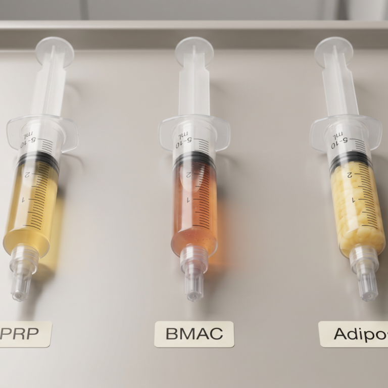
Before discussing technique, clinicians should be clear about the regulatory status of the products they inject. PRP preparation systems are cleared by the U.S. Food and Drug Administration under the 510(k) pathway for bone graft handling. Clinicians process autologous blood into platelet concentrate using these cleared devices. The FDA has not approved PRP for specific disease indications such as knee osteoarthritis, tendinopathy, hair loss, or aesthetic applications. All of these uses are considered off-label.
Off-label means the FDA has not reviewed safety or efficacy data for that specific indication. Clinicians may still use cleared devices off-label in the exercise of clinical judgment. Patients must be informed that the use is off-label, that evidence is mixed, and that outcomes are not guaranteed. Documentation of that conversation belongs in the chart.
Other regenerative products require careful regulatory framing. Adipose stromal vascular fraction (SVF) preparations that involve enzymatic processing are generally considered unapproved 351 biologics and should not be marketed as treatments. Exosome products have no FDA-approved indications, and the agency has issued consumer alerts about unapproved stem cell and exosome products. Cord blood hematopoietic progenitor cells are approved for specific blood disorders and do not apply to musculoskeletal injections. Clinicians who want to understand how these product categories intersect with day-to-day practice should consult current FDA guidance and their own legal counsel before offering any of them.
Safety considerations and complication prevention
The safety case for ultrasound guidance rests on two ideas. First, imaging helps clinicians avoid structures they should not hit. Second, seeing the needle tip changes how they react when something looks wrong. Both matter for regenerative injections, where volumes can be higher and the target is often close to neurovascular structures.
Several specific harm mechanisms are documented in the interventional pain and regional anesthesia literature. Vascular puncture can produce hematomas that compress nearby nerves. Intraneural or intrafascicular injection can cause mechanical, pressure, and chemical nerve injury. High injection pressures during deep blocks can drive injectate into unintended spaces.
Training should address each of these failure modes directly. Operators need to recognize the sonographic appearance of arteries, veins, and nerves. They must hold the probe steady enough to track the needle tip at all times. They also need to apply hydrodissection, which means separating tissue planes with saline or local anesthetic under imaging, to open safe corridors. Finally, they should pay attention to injection pressure as a warning signal. Most of these skills are developed through supervised repetition rather than didactic study alone.
What this means in practice: Ultrasound guidance does not make injections risk-free. It gives trained operators tools to identify and avoid specific harms, but those tools only help when the operator knows what to look for and what to do about it.
Fictional case example: the cost of the learning curve
The following is a fictional composite clinical scenario created for educational purposes. It does not describe a real patient.
A 54-year-old family medicine physician attends an introductory weekend course on PRP for musculoskeletal indications. She returns to her practice excited to offer knee PRP to appropriate patients. She buys a PRP kit and a portable ultrasound unit. Her first six patients include three with mild-to-moderate knee osteoarthritis and three with rotator cuff tendinopathy.
After six weeks, two of the knee patients report no change, and one patient with rotator cuff tendinopathy describes new numbness in the lateral arm that resolves over several days. Concerned, she reviews her injection videos. On two of the knee cases, her needle appears to sit just outside the joint capsule. On the rotator cuff case, her needle path crossed closer to the axillary nerve region than she realized in the moment.
She pauses her PRP program, enrolls in a structured musculoskeletal ultrasound course with supervised live-patient training, and spends three months practicing scanning and needle placement under faculty observation before resuming. The lesson is not that she was negligent. The lesson is that a weekend of didactics is not enough to build needle-tracking competence. The most dangerous moment in procedural practice is the one when a clinician believes they can see more than they actually can.
Key lesson: Structured, supervised training on live patients is the shortest safe path from basic familiarity to reliable technique. Skipping that step puts patients and licenses at risk.
Who should learn these techniques
Ultrasound-guided injection is within scope for many types of licensed clinicians when local regulations allow. This includes physicians in sports medicine, physical medicine and rehabilitation, orthopedics, family medicine, pain medicine, emergency medicine, and aesthetics. Advanced practice providers working in musculoskeletal care may also perform these procedures under appropriate supervision and scope rules. Scope of practice varies by state and by credentialing body, so each clinician needs to confirm that procedural ultrasound and the specific regenerative injection are permitted under their license and institutional policy.
Beyond licensure, a good candidate for training sees enough musculoskeletal complaints to build repetition. They also have time to practice scanning between cases. Most important, they accept that competence takes months rather than weeks. Clinicians who plan to offer only occasional injections should think carefully about whether intermittent practice will maintain the skill safely.
The learning curve and why structured training matters
Musculoskeletal ultrasound is often described as technically challenging with a steep learning curve. Image acquisition depends on probe handling, and interpretation depends on knowing sonographic anatomy and recognizing common artifacts. Neither skill develops quickly, and both atrophy without regular use.
The training landscape in the United States adds to the challenge. The title sonographer is unregulated in most contexts, which means clinicians can legally perform ultrasound without formal coursework. That freedom creates medicolegal risk, particularly for solo practitioners who do not have peers to review images with them. There is no single mandatory national curriculum for physicians, although the AMSSM has published a recommended sports ultrasound curriculum for fellowships and several certifying bodies offer voluntary credentials.
Commercial musculoskeletal training providers commonly describe foundational programs that span two to three months of part-time study with hands-on practice. True procedural competence tends to develop over longer periods, with supervised scanning and injection volume accumulating alongside didactic work. Clinicians who approach ultrasound training as a one-time event rather than a phased skill build tend to plateau. They reach a level where they can acquire images but cannot reliably interpret or act on them. Programs that pair didactic instruction with supervised procedural volume under credentialed physician faculty produce more reliable technique than standalone lectures or weekend workshops.
What this means in practice: Competence is a function of supervised repetition, not course duration alone. A well-designed training pathway combines didactic instruction with repeated live-patient practice and faculty feedback.
Core competencies a quality training program should cover
A training program that prepares clinicians to offer ultrasound-guided regenerative injections should cover a defined set of competencies. Synthesizing guidance from the AAPM&R position statement, the AMSSM curriculum, and the broader interventional MSK ultrasound literature yields the following core domains.
Operators need a working understanding of ultrasound physics and knobology, including frequency, gain, depth, and focal zone settings. They need to choose probes appropriately for the anatomic target. They need to recognize the sonographic appearance of normal and pathologic muscle, tendon, ligament, bursa, nerve, and vessel. They need to identify common artifacts such as anisotropy, posterior acoustic enhancement, and reverberation, because artifacts cause real misinterpretations during injections.
On the procedural side, operators need to develop needle visualization technique using both in-plane and out-of-plane approaches. They should practice hydrodissection when compartment identification is difficult. They should also build awareness of injection pressure as a warning for intraneural or malpositioned needles. Sterile technique, probe-cover protocols, and image documentation all belong in a complete program. Image documentation is particularly important, because a saved pre-injection and post-injection clip serves both clinical and medicolegal purposes.
Finally, a regenerative-focused program should address patient selection, informed consent, and off-label disclosure specific to products like PRP. The science of PRP itself is relevant, because platelet biology, preparation parameters, and concentration thresholds have been linked in some studies to outcome differences, even if the evidence base is heterogeneous. A clinician who understands why a given preparation matters is better positioned to counsel patients honestly and to interpret outcome variability in their own practice.
Credentialing pathways for clinicians
There is no single required credential for physicians or advanced practice providers who perform musculoskeletal ultrasound in the United States. Several voluntary pathways exist, and they vary in scope and recognition.
The Registered in Musculoskeletal Sonography (RMSK) credential, offered by the Alliance for Physician Certification and Advancement, is the most commonly cited physician and advanced practice provider certification in this space. Eligibility requires a valid medical license in good standing plus a passing score on the written musculoskeletal examination. Applicants must also document at least 150 diagnostic musculoskeletal ultrasound studies completed on actual patients within the preceding 36 months. That clinical volume requirement is a meaningful prerequisite and reinforces the point that the credential reflects real scanning experience, not coursework alone. A companion credential for sonographers, the Registered Musculoskeletal Sonographer (RMSKS), is offered through ARDMS within the same Inteleos credentialing family. Both credentials are voluntary and serve different practice roles.
Beyond formal credentials, society-level curricula from AMSSM and AAPM&R provide recognized frameworks for fellowship training and continuing medical education. A clinician building a training pathway should combine at least one recognized curriculum or credential with substantial supervised procedural volume. Clinicians should also remember that the marketing phrase board-certified in regenerative medicine does not refer to an American Board of Medical Specialties board. Accuracy in how you describe your own credentials matters for both compliance and patient trust.
Practice integration and cost considerations
Integrating ultrasound guidance into a regenerative practice changes workflow in several ways. Diagnostic musculoskeletal ultrasound is significantly less expensive than magnetic resonance imaging, and appropriate use in the office can reduce downstream imaging costs. Portable point-of-care units allow diagnosis and guided intervention in the same visit, which is convenient for patients and for clinic throughput.
Guided injections do take longer than blind injections. A cadaveric study of shoulder injections reported average procedure times of roughly 166 seconds for ultrasound-guided injections versus 52 seconds for freehand injections. For most regenerative cases this trade-off is acceptable, because the accuracy gain outweighs the extra time, but clinicians need to budget for it when scheduling.
Coding and coverage for ultrasound guidance vary by payer and by indication. Many regenerative injections are offered off-label and often on a cash-pay basis. Clinicians should work with coders and legal counsel to structure transparent fee arrangements. These arrangements should avoid mixing covered and non-covered services in misleading ways. Patients should receive clear written information about what is covered, what is off-label, and what is out-of-pocket before any procedure.
Frequently asked questions
How long does it realistically take to become competent in musculoskeletal ultrasound for regenerative injections?
Competence develops over months rather than weeks. Many structured training programs span two to three months of part-time foundational work, and procedural confidence typically builds across the following months with supervised live-patient practice. Individual timelines vary based on prior imaging experience, practice volume, and access to structured feedback.
Is ultrasound guidance required for PRP injections?
Ultrasound guidance is not universally required by regulation, but it is strongly supported by the accuracy literature for most joints and soft tissues, and it is increasingly expected by professional societies and patients. For small joints, deep targets, and anatomically complex structures, many clinicians consider guidance the standard of care in their own practice.
Does higher placement accuracy translate into better patient outcomes?
Accuracy and clinical outcomes are related but not identical. At several sites, including the subacromial space and the intra-articular knee, ultrasound guidance improves both accuracy and short-term pain outcomes. At other sites, such as the first carpometacarpal joint, accuracy gains have not translated into clear clinical superiority across all outcome measures. Honest counseling should reflect this nuance.
What is the most common safety issue with untrained operators?
Failure to reliably track the needle tip is the recurring issue. When operators lose visualization, the margin for error shrinks, and injection into or near a nerve, vessel, or unintended compartment becomes more likely. Supervised practice with deliberate attention to needle tracking is the main countermeasure.
How should I counsel a patient who has already had an unsuccessful PRP injection elsewhere?
Begin by asking about the prior preparation, whether ultrasound was used, and what outcomes were discussed in advance. Many unsuccessful cases reflect a combination of unclear expectations, heterogeneous preparation, and variable placement. A second attempt may be reasonable in some scenarios, but patients deserve a frank conversation about evidence, off-label status, and the possibility of no meaningful benefit before proceeding.
Do I need a separate credential to bill for ultrasound guidance?
Billing and credentialing requirements vary by payer, institution, and state. Some payers expect documented training or specific credentials before reimbursing for guidance codes. Clinicians should confirm requirements with their coding staff, their malpractice carrier, and their institution before building a program around guided procedures.
How do I counsel patients about off-label PRP use?
Explain that PRP preparation systems are FDA-cleared for bone graft handling and that use for knee osteoarthritis, tendinopathy, hair restoration, and aesthetic indications is off-label. Clarify that off-label use is legal but means the FDA has not reviewed safety or efficacy data for that specific indication. Document the conversation, including expected response rates, possible adverse events, and the absence of outcome guarantees.
When should I refer a patient rather than manage them in-house?
Refer when the diagnosis is uncertain, when the target anatomy is outside your training, when prior procedures have not resolved the issue, or when the patient has comorbidities that raise procedural risk. A thoughtful referral strengthens trust and protects patients from preventable harm.
Key takeaways
- Ultrasound-guided injections consistently improve needle placement accuracy compared with landmark-guided injections, with the strongest evidence at the shoulder, knee, small joints, and carpal tunnel.
- For regenerative injections such as PRP, accurate delivery is a prerequisite for any biologic effect, not a guarantee of patient outcomes. PRP is FDA-cleared only for bone graft handling and used off-label for musculoskeletal and aesthetic indications.
- Clinical outcome advantages for ultrasound guidance are strong at some sites and mixed at others; honest counseling reflects that nuance rather than overstating benefit.
- Musculoskeletal ultrasound has a steep learning curve, and competence develops over months of supervised practice rather than during a single weekend course.
- Core competencies include ultrasound physics, sonographic anatomy, artifact recognition, in-plane and out-of-plane needle tracking, hydrodissection, injection-pressure awareness, and image documentation.
- Voluntary credentials such as RMSK provide recognized frameworks, but there is no single required national credential for U.S. physicians.
- Practice integration requires attention to workflow, scheduling, coding, and transparent cost disclosure for off-label procedures.
- Structured, live-patient training with faculty feedback is the shortest safe path from basic familiarity to reliable procedural skill.
RMA Disclosure
Regenerative Medicine Academy (RMA) is an education company that provides clinician training in some of the techniques discussed in this article. This content is educational and does not constitute a claim of clinical superiority, a guarantee of outcomes, or a substitute for individual clinical judgment.
Build the skills your patients expect
Ultrasound guidance is changing what safe, defensible regenerative practice looks like. If you are ready to develop procedural skills under faculty observation with live patients, explore RMA’s hands-on regenerative medicine training courses. Structured e-learning coursework supports licensed clinicians who want to integrate these techniques responsibly. Competence is built through supervised repetition, and the right training environment makes that possible.